




Slit Lamp Eye Exam: What It Shows, Why It Matters, and What to Expect
Your eyes can develop changes long before you notice blurry vision, pain, or discomfort. A slit lamp eye exam allows an ophthalmologist to examine the front and back structures of the eye under high magnification, helping detect problems at an early stage when treatment is often most effective.
Many patients visiting an eye clinic in Flushing, Queens, New York City ask whether this test is painful or why it is performed even when their vision seems normal. The answer is simple: many eye conditions progress silently. A slit lamp examination gives your ophthalmologist a detailed view of delicate eye tissues that cannot be evaluated with a standard vision chart alone.
At LeeVision, Board-Certified Ophthalmologist Dr. David H. Lee, MD uses advanced diagnostic technology as part of a comprehensive eye examination to evaluate eye health, identify early disease, and create personalized treatment recommendations based on each patient’s needs.
Why a Slit Lamp Eye Exam Is One of the Most Important Eye Tests
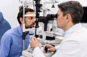
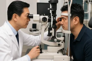
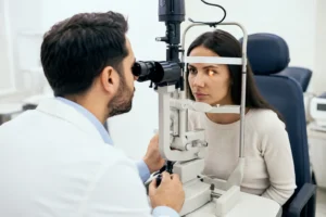
A slit lamp is a specialized microscope combined with a bright, narrow beam of light. By changing the angle, width, and intensity of the light, your ophthalmologist can examine different layers of the eye in remarkable detail.
Unlike a routine vision screening that measures how clearly you see letters on a chart, a slit lamp eye exam evaluates the actual health of your eyes.
It helps detect changes involving:
- Cornea
- Conjunctiva
- Iris
- Lens
- Eyelids
- Tear film
- Anterior chamber
- Retina (when combined with dilating eye drops and special lenses)
- Optic nerve
Because many eye diseases begin with subtle structural changes rather than noticeable symptoms, this examination plays an essential role in preventive eye care.
Who Should Have a Slit Lamp Eye Exam?
Although the test is commonly performed during comprehensive eye exams, some people benefit from it more frequently.
Your ophthalmologist may recommend regular slit lamp examinations if you:
- Wear contact lensesUnderstanding the Waggoner Color Vision Test
- Have diabetes
- Have high blood pressure
- Experience dry eyes
- Notice blurry vision
- Have eye redness or irritation
- Suffer an eye injury
- Have a family history of glaucoma
- Are preparing for cataract or LASIK surgery
- Are over age 40 and receiving routine preventive eye care
In a busy community like Flushing, Queens NYC, many adults spend long hours using computers, smartphones, and digital devices. While digital eye strain itself does not damage the eye, prolonged screen use can worsen dry eye symptoms that are often easier to evaluate during a slit lamp examination.
What Can a Slit Lamp Eye Exam Detect?
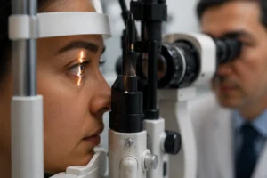
One reason ophthalmologists rely on this examination is that it provides detailed information about multiple eye structures during a single appointment.
Common conditions identified include:
Dry Eye Disease
The slit lamp allows your doctor to examine the tear film, eyelids, and meibomian glands that produce the oily layer of tears.
Patients often describe symptoms such as:
- Burning eyes
- Grittiness
- Fluctuating vision
- Excessive tearing
- Eye fatigue after screen use
These symptoms may seem minor but can significantly affect daily comfort and vision quality.
Cataracts
As we age, the eye’s natural lens gradually becomes cloudy.
A slit lamp eye exam helps determine:
- Whether cataracts are present
- Their location
- Their severity
- Whether they are likely causing your vision changes
Many patients are surprised to learn that cataracts develop slowly over several years before surgery becomes necessary.
Corneal Disorders
The cornea is the clear front window of the eye.
Using the slit lamp, an ophthalmologist can detect:
- Corneal scratches
- Infections
- Swelling
- Corneal ulcers
- Keratoconus
- Degenerative changes
- Foreign bodies
Because the cornea contains many nerve endings, even small injuries can cause significant discomfort, making early diagnosis important.
Glaucoma Evaluation
Although measuring eye pressure is an important part of glaucoma screening, pressure alone does not diagnose glaucoma.
A slit lamp examination helps evaluate:
- The drainage angle
- Corneal thickness
- Anterior chamber depth
- Signs that may increase glaucoma risk
When combined with additional diagnostic testing, it provides valuable information for preserving long-term vision.
Eye Allergies and Inflammation
Many patients assume itchy or red eyes are simply seasonal allergies.
However, a slit lamp examination can distinguish between:
- Allergic conjunctivitis
- Viral infections
- Bacterial infections
- Blepharitis
- Uveitis
- Dry eye disease
Since these conditions often produce similar symptoms, an accurate diagnosis helps guide appropriate treatment rather than relying on guesswork or over-the-counter drops alone.
What Happens During a Slit Lamp Eye Exam?
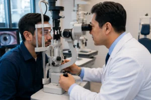
The examination is straightforward and typically takes
only a few minutes.
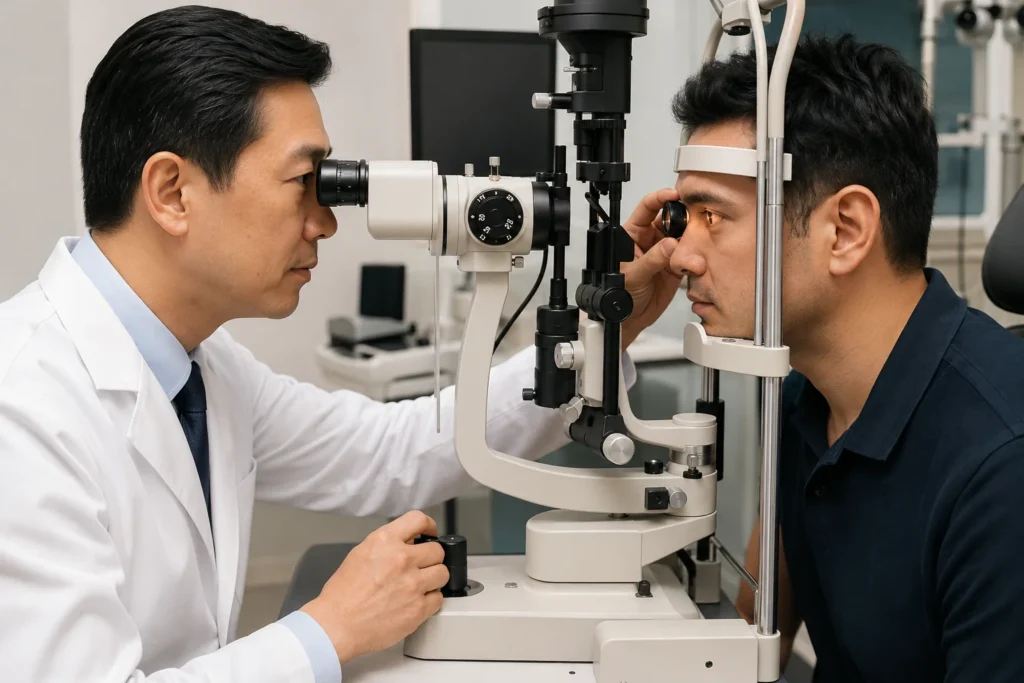
First, you’ll sit comfortably in front of the slit lamp microscope. You’ll place your chin on a chin rest and your forehead against a support to help keep your head steady.
Your ophthalmologist then shines a narrow beam of light into different areas of your eyes while viewing them through high-powered magnification.
Depending on your symptoms or medical history, additional steps may include:
- Applying fluorescein dye to evaluate the cornea
- Measuring eye pressure
- Dilating the pupils to examine the retina and optic nerve
- Using specialized lenses to inspect deeper eye structures
Patients often worry that the bright light may be uncomfortable, but the test is generally painless. You may briefly notice increased sensitivity to light, especially if dilation is performed.
Why Early Detection Matters More Than Most People Realize
One of the biggest misconceptions patients have is believing that good vision automatically means healthy eyes.
In reality, several serious conditions—including glaucoma, diabetic eye disease, and early cataracts—can progress without causing obvious symptoms.
A slit lamp eye exam allows ophthalmologists to identify subtle changes before permanent vision loss occurs, giving patients more treatment options and better long-term outcomes.
Slit Lamp Eye Exam vs. a Regular Eye Exam
Many patients assume that a vision test and a slit lamp eye exam are the same thing. While both are important, they evaluate different aspects of your eye health.
| Regular Vision Test | Slit Lamp Eye Exam |
|---|---|
| Measures how clearly you see | Examines the health of eye structures |
| Checks your glasses or contact lens prescription | Detects eye diseases and abnormalities |
| Uses a vision chart | Uses a high-powered binocular microscope |
| Focuses on visual acuity | Focuses on eye health and disease detection |
| Usually takes only a few minutes | Often performed as part of a comprehensive eye exam |
Think of it this way: a vision chart tells your ophthalmologist how well you see, while a slit lamp eye exam helps explain why your vision may have changed or whether there are early signs of disease.
Is a Slit Lamp Eye Exam Safe?
Yes. A slit lamp eye exam is considered one of the safest and most commonly performed diagnostic procedures in ophthalmology.
The microscope never touches your eye. The examination uses focused light and magnification to allow your ophthalmologist to inspect the eye’s structures in detail.
Some patients may experience temporary light sensitivity, especially if dilating eye drops are used. If your pupils are dilated, your near vision may remain blurry for several hours, and sunglasses can make you more comfortable when going outdoors.
There are no lasting side effects from the slit lamp examination itself.
Can a Slit Lamp Eye Exam Detect Problems Behind the Eye?
Yes—but this depends on whether your pupils are dilated.
Without dilation, your ophthalmologist can thoroughly examine the front part of the eye. When dilation is added, specialized lenses used with the slit lamp provide a detailed view of the retina, macula, optic nerve, and retinal blood vessels.
This is particularly important for people with:
- Diabetes
- High blood pressure
- Family history of retinal disease
- High myopia (severe nearsightedness)
- Flashes of light or new floaters
- Sudden vision changes
A dilated slit lamp examination often reveals problems long before patients notice symptoms.
Understanding the Waggoner Color Vision Test
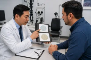
Color vision is an essential part of many comprehensive eye examinations, especially when patients report difficulty distinguishing certain colors or when color perception is important for work or daily activities.
The Waggoner Color Vision Test is a modern screening tool designed to evaluate color vision deficiencies more consistently than many traditional methods.
Unlike older printed color plates that can fade over time or vary with room lighting, newer Waggoner testing systems are designed to provide more standardized testing conditions, improving the reliability of results.
An ophthalmologist may recommend this test if you:
- Have difficulty identifying certain colors
- Have a family history of color blindness
- Need occupational vision testing
- Are undergoing a neurological or retinal evaluation
- Require documentation for school or employment
The test is quick, painless, and requires patients to identify numbers, symbols, or patterns displayed in specially designed color images.
What Can the Waggoner Color Vision Test Detect?
The test helps identify different types of color vision deficiencies, including:
- Red-green color deficiency
- Blue-yellow color deficiency
- Partial color vision loss
- Complete color blindness (rare)
Although most color vision deficiencies are inherited, changes in color perception can also occur because of certain eye diseases, optic nerve disorders, retinal conditions, or medication side effects. If color vision changes suddenly, it should always be evaluated promptly.
Understanding the FAA Color Vision Test
For individuals pursuing a career in aviation, color vision is more than a convenience—it is an important safety requirement.
The FAA Color Vision Test helps determine whether pilots can accurately recognize aviation signal lights, cockpit displays, runway lighting, navigation charts, and other color-dependent information necessary for safe flight operations.
Not everyone applying for pilot certification undergoes the exact same test. The Federal Aviation Administration accepts several approved methods of evaluating color vision, and the specific test may vary depending on the aviation medical examiner and current FAA guidelines.
Many applicants are concerned after failing an initial color vision screening. However, failing one screening test does not always mean a person is permanently disqualified. In some situations, additional FAA-approved testing may be available to better assess functional color vision.
If you are preparing for an aviation medical examination, discussing your vision history with an experienced ophthalmologist beforehand can help you understand what to expect.
Can a Slit Lamp Eye Exam Diagnose Color Blindness?

A slit lamp eye exam and a color vision test evaluate different aspects of eye health.
The slit lamp examines the physical structures of the eye, while tests such as the Waggoner Color Vision Test or FAA Color Vision Test evaluate how your eyes and brain perceive colors.
However, these tests often complement one another. If an ophthalmologist notices signs of retinal disease or optic nerve damage during a slit lamp examination, additional color vision testing may help determine whether those conditions are affecting color perception.
Common Misconceptions About Slit Lamp Eye Exams
Many people postpone eye examinations because of misunderstandings about the test. Here are some of the most common myths.”If I can see clearly, I don’t need the test.”
Many eye diseases—including glaucoma, diabetic eye disease, and early cataracts—can develop without affecting vision initially. A slit lamp eye exam helps detect these problems before symptoms appear.
“The bright light can damage my eyes.”
The light used during the examination is carefully controlled and safe for routine clinical use. Temporary brightness is normal, but it does not harm healthy eyes.
“The examination is painful.”
Most patients experience no pain. The only mild discomfort is brief light sensitivity, particularly if your pupils are dilated.
“Only older adults need a slit lamp examination.”
People of all ages may benefit from this examination. Children, contact lens wearers, office workers with digital eye strain, athletes, and individuals with chronic health conditions may all require slit lamp evaluations at different stages of life.
When Should You Schedule a Comprehensive Eye Examination?
While routine eye exams are important, certain symptoms should never be ignored.
Arrange an eye evaluation promptly if you notice:
- Sudden blurred vision
- Persistent eye pain
- New flashes of light
- A sudden increase in floaters
- Double vision
- Eye injuries
- Ongoing redness that does not improve
- Frequent headaches associated with vision changes
- Difficulty seeing at night
- Changes in color perception
Early evaluation often leads to earlier treatment, helping protect long-term vision and reduce the risk of permanent eye damage.
Why Comprehensive Eye Care Matters
A slit lamp eye exam is far more than a routine part of an eye checkup. It provides a detailed view of structures that cannot be assessed through a vision chart alone, making it one of the most valuable tools for detecting eye disease at an early stage.
When combined with specialized evaluations such as the Waggoner Color Vision Test and, when appropriate, the FAA Color Vision Test, ophthalmologists gain a more complete understanding of both the health and function of your eyes.
For individuals in Flushing, Queens, New York City, regular comprehensive eye examinations are especially important in today’s screen-focused lifestyle, where digital eye strain, dry eye disease, diabetes, and age-related eye conditions are increasingly common. Early diagnosis, personalized care, and ongoing monitoring remain the best strategies for protecting vision throughout life.
Frequently Asked Questions
What is a slit lamp eye exam?
A slit lamp eye exam is a detailed eye examination that uses a specialized microscope and a narrow beam of light to evaluate the front and, when necessary, the back of the eye. It helps detect cataracts, glaucoma, dry eye disease, corneal injuries, infections, and other eye conditions before noticeable symptoms develop.
Why is this examination important?
This examination allows an ophthalmologist to identify early signs of eye disease that may not affect your vision initially. Detecting problems early often leads to more effective treatment and better long-term eye health.
How long does the procedure take?
The test itself usually takes between 5 and 10 minutes. If pupil dilation or additional imaging is needed, your complete appointment may last between 30 and 90 minutes.
Is the test painful?
No. The procedure is painless and non-invasive. You may notice a bright light during the examination, but it does not damage the eyes. If dilation is performed, temporary light sensitivity is normal.
Do I need to prepare before my appointment?
Most patients do not need any special preparation. If your eyes will be dilated, bringing sunglasses is recommended since your vision may remain blurry and light-sensitive for a few hours afterward.
Can this examination detect glaucoma?
Yes. It helps your ophthalmologist evaluate important structures such as the drainage angle, cornea, and anterior chamber. Combined with pressure testing and optic nerve assessment, it plays an important role in glaucoma detection.
Can it detect cataracts?
Yes. This microscope-based examination allows your ophthalmologist to see whether the natural lens has become cloudy and determine how much the cataract is affecting your vision.
Can it diagnose dry eye disease?
Yes. The examination provides a close view of the tear film, eyelids, and meibomian glands, helping identify the cause of dry eye symptoms and guide appropriate treatment.
Can retinal diseases be detected?
Yes. When your pupils are dilated, specialized lenses allow the ophthalmologist to examine the retina, macula, and optic nerve for diabetic retinopathy, macular degeneration, retinal tears, and other conditions.
Does this test check my glasses prescription?
No. Your eyeglass or contact lens prescription is measured separately during a vision test. This examination focuses on evaluating the health of your eyes rather than determining lens power.
Who should have this examination?
It is recommended for adults and children who have vision changes, diabetes, glaucoma risk factors, dry eye symptoms, eye injuries, or wear contact lenses. Routine eye examinations are also advised as part of preventive eye care.
Is it safe for children?
Yes. Ophthalmologists frequently perform this examination on children when evaluating infections, eye injuries, congenital conditions, or unexplained vision problems.
What is the Waggoner Color Vision Test?
The Waggoner Color Vision Test is an advanced screening method used to identify inherited or acquired color vision deficiencies. It provides standardized testing conditions that improve the consistency of color vision evaluation.
Who should take the Waggoner Color Vision Test?
This assessment may be recommended for students, pilots, electricians, healthcare professionals, designers, and individuals with suspected color vision deficiency or certain retinal and optic nerve disorders.
What is the FAA Color Vision Test?
The FAA Color Vision Test is used during aviation medical certification to confirm that pilots can accurately distinguish aviation signal lights and color-coded flight information required for safe flying.
What happens if I fail the FAA Color Vision Test?
Failing an initial screening does not always prevent certification. Depending on current FAA guidelines, additional FAA-approved testing options may be available through an Aviation Medical Examiner.
Is the Waggoner Color Vision Test the same as the FAA Color Vision Test?
No. The Waggoner Color Vision Test is commonly used in clinical eye care, while the FAA Color Vision Test follows aviation-specific certification standards. Although both evaluate color perception, they serve different purposes.
Can this examination diagnose color blindness?
No. Color blindness is diagnosed using dedicated color vision assessments, such as the Waggoner Color Vision Test or approved FAA color vision evaluations, rather than the microscope examination itself.
How often should adults have this examination?
The recommended schedule depends on your age, overall eye health, and medical history. People with diabetes, glaucoma, cataracts, or retinal disease usually require more frequent comprehensive eye examinations.
Where can I receive a comprehensive eye examination in Flushing, Queens, NYC?
Patients looking for advanced eye care in Flushing, Queens, New York City should choose a practice with modern diagnostic technology and a board-certified ophthalmologist. At LeeVision, comprehensive eye examinations are personalized based on each patient’s vision needs and overall eye health.